Identify Mushrooms by Photo
Dr. MycoTek asks the questions a field mycologist asks — what shape is the cap, gills or pores underneath, any ring on the stem, what is it growing from? Then explains what each answer means for the ID.
No credit card required · Free forever tier
Or jump into the live tools
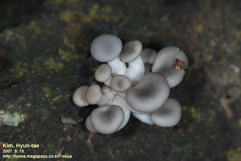
© pintail (iNaturalist, cc-by)
How a Mycologist Reads a Mushroom Photo
When a mycologist looks at a mushroom photo, they do not try to match it to a remembered image. They read a sequence of diagnostic features in a deliberate order: cap shape and surface first, then the underside (gills, pores, or teeth), then the stem (proportions, ring, base), then the ecological context (substrate, nearby trees, season). Dr. MycoTek follows the same sequence. When you upload a photo, it does not return a single confident answer — it asks the questions that narrow the field, explains what each feature tells us, and builds the identification with you step by step. The goal is not just an answer but an understanding of why that answer is supported.
Taking Better Photos for Identification
The single most impactful thing you can do is take multiple photos from different angles. At minimum, capture: a top-down view of the cap showing colour and texture, an underside view showing the gill or pore structure clearly, a side profile showing the stem and overall proportions, and a close-up of the stem base (gently dig around the base to expose any volva or bulb). Natural daylight produces the most accurate colours — flash photography can wash out subtle colour differences that are critical for identification. Include a coin or your finger for scale, and if possible, photograph the mushroom in situ before picking it, so the habitat context is preserved.
The Diagnostic Features That Matter Most
Dr. MycoTek works through several diagnostic categories from your image. Cap morphology is assessed first: is the cap convex, flat, funnel-shaped, or conical? Is the margin smooth, lined, or ragged? Surface texture provides additional clues — the sticky cap of a Suillus bolete versus the dry, scaly cap of a Pholiota are immediately distinctive. Underneath the cap, the hymenium type (gills, pores, teeth, or false ridges) is the most critical structural feature. On the stem, the presence or absence of a ring and the shape of the base — bulbous, tapered, or with a cup-like volva — are essential, especially for separating edible species from dangerous Amanita relatives.
What a Photo Cannot Show You
Even with excellent photographs, certain features cannot be assessed visually. Spore colour — one of the most reliable diagnostic features — requires a spore print on paper. Odour is often highly diagnostic (anise smell in Clitocybe odora, bleach smell in Mycena, mealy smell in many Entoloma). Chemical reactions like the flesh of certain boletes staining blue when cut require physical interaction with the specimen. Dr. MycoTek will tell you when these non-visual tests would significantly improve confidence and provide clear instructions for performing them. The photo starts the conversation; these additional checks finish it.
Common Photo Identification Mistakes
The most frequent mistake is photographing only the top of the cap. The underside (gills or pores) and the stem base are often more important than the cap surface for accurate identification. Another common error is photographing mushrooms that are too old — colours fade, gills darken, and structural features deteriorate as mushrooms age. Removing the mushroom from its substrate before photographing also eliminates critical habitat information. Always note whether it was growing from soil, dead wood, living wood, dung, or leaf litter — substrate is one of the first things a mycologist asks about.
Mobile Photography Tips
Most mushroom photos come from smartphones, and modern phone cameras are more than adequate. Use macro mode to capture fine details like gill structure. Tap to focus on the specific feature you want sharp — autofocus often locks onto the wrong plane when photographing small subjects at close range. Avoid digital zoom; get physically closer. If the mushroom is in deep shade, hold a piece of white paper nearby to bounce light onto the underside without creating harsh shadows. Wet mushrooms are tricky — water droplets obscure surface details, so gently blot excess moisture if you can.
When to Ask for Additional Checks
Dr. MycoTek will explicitly tell you when a photo is not enough. This commonly occurs with small brown mushrooms (LBMs — Little Brown Mushrooms), white Agaricus-like species that could be confused with Death Caps, and boletes where pore colour and staining reactions are essential. In these cases, it provides a shortlist of candidate species and tells you exactly which additional tests to perform — spore print, chemical tests, or microscopy. It may also recommend posting the specimen to a local mycological society or iNaturalist for community verification. The clearest outcome is when you can say: I checked these features, and they match only one candidate.
What You Get
See It In Action
Real photos from the community
Curated commercial-license observations from Mushroom Observer.


Frequently Asked Questions
Related Topics
Ready to Get Expert Help?
Dr. MycoTek is free to start. No credit card required.
Trained on 12 million words of real grower knowledge. 80+ species. 4,400+ reference photos.
No credit card required · Free forever tier